learn unique, effective techniques, and how to create too much business!
muscle box 4 - front of hips and legs and reflexology
Almost finished! Just a few more muscles to learn.
You could search in Internet, read blogs, and watch lots of online videos, OR you can take a short cut and get some very good anatomy education RIGHT HERE. We have done a LOT of leg work to put this valuable information in one place. PLUS, a lot of this comes from many years of getting results for thousands of clients.
The "Muscle Box" - This is a list of muscles we learned to target as we perform therapy. If you learn this list, you'll have a good basic understanding of many of the tight muscles that can affect your clients. We have been getting performance feedback from clients since 1989 so we know this list is a vital key to your capability to help your clients. You will see:
(a) The muscle name and picture,
(b) The action of the muscle
(c) The attachment points (We sometimes use generic descriptions.) and
(d) A "nickname" you can you use in your progress notes.
(e) "Why this is important section". Extra information to help your application of this learning.
Note: Even though this course is "basic anatomy", it is the building block of both your ability to perform our therapy style, AND your ability to communicate what you are doing.

John at Houston's Memorial Park
anterior hips and legs

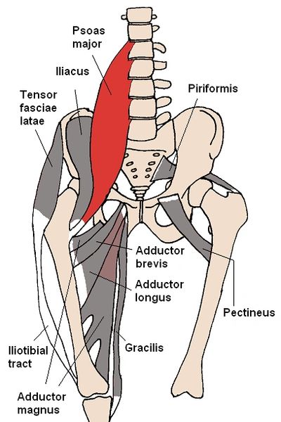
1. Psoas
Beth ohara / CC BY-SA (http://creativecommons.org/licenses/by-sa/3.0/)
Action
The psoas major is a long fusiform (wide in middle and tapers at the ends) muscle located in the lateral lumbar region between the vertebral column and the brim of the lesser pelvis. It joins the iliacus muscle to form the iliopsoas
Action: Flexion in the hip joint (like raising your knee, or a sit up motion)
Attachments
The Psoas runs from the FRONT of the lumbar spine "like an upside down V" to the medial upper femur.
Origin: Transverse processes of T12-L4 and the lateral aspects of the discs between them (front side of the lower spine)
Insertion: The lesser trochanter of the femur (inside of top of leg bone)
Nickname
psoas
Why is this important?
When you work on the hip flexors, you usually cover both the Psoas and Iliacus. You'll want to note if you find one muscle to be tighter than the other. We have several different ways to work on the hip flexors. ALSO, we will teach you what spot on the low back indicates Psoas tightness on the front.

2. Illiacus
Beth ohara / CC BY-SA (http://creativecommons.org/licenses/by-sa/3.0/)
Action
The iliacus is a flat, triangular muscle which fills the iliac fossa. It forms the lateral portion of iliopsoas, providing
Action: flexion of the thigh and lower limb (like a sit up motion)
Attachments
Origins: upper two-third of the iliac fossa (inside the pelvis)
Insertion: base of the lesser trochanter of femur (inside top of the leg bone)
Nickname
Iliacus
Why is this important?
When you work on the hip flexors, you usually cover both the Psoas and Iliacus. You'll want to note if you find one muscle to be tighter than the other. We have several different ways to work on the hip flexors.

3. Pectineus
Beth ohara / CC BY-SA (http://creativecommons.org/licenses/by-sa/3.0/)
Action
The pectineus muscle is a flat, quadrangular muscle, situated at the anterior part of the upper and medial aspect of the thigh. The pectineus muscle is the most anterior adductor of the hip.
Action: (1) adduct and internally rotate the thigh but its primary function is (2) hip flexion
Attachments
Origin: Pectineal line of the pubic bone
Insertion: Pectineal line of the femur
Nickname
pectineus
Why is this important?
When the pectineus is tight the client usually points right to this area. We will teach you some therapy techniques in the Tool Box.
Site Content

4. Adductor group
Beth ohara / CC BY-SA (http://creativecommons.org/licenses/by-sa/3.0/)
Action
The adductor muscles of the hip are a group of muscles mostly used for bringing the thighs together.
Action: Adduction (inward movement) of hip
Attachments
Origin: Pubis (bottom of the Pelvis)
Insertion: Femur and Tibia (both upper AND lower legs)
Nickname
a) Add
b) Location i.e. proximal Add or distal Add
Why is this important?
(a) If the Adductors are part of the problem we have several body positions you will want to use.
(b) We will teach you how to get the client to show you the key "pain" location that indicates Adductor tightness.

5. Gracilus
Beth ohara / CC BY-SA (http://creativecommons.org/licenses/by-sa/3.0/)
Action
The gracilis muscle is the most superficial muscle on the medial side of the thigh.
Action: (1) flexes, (2) medially rotates, and (3) adducts the hip
Attachments
Origin: ischiopubic ramus (lower pelvis)
Insertion: tibia (lower leg bone)
Nickname
Gracilus
Why is this important?
Although this muscle is relatively small, it is important to palpate and discover if it is part of the client's problem.

6. Sartorius
modified by Uwe Gille / Public domain
Action
The sartorius muscle is the longest muscle in the human body. It is a long, thin, superficial muscle that runs down the length of the thigh in the anterior compartment.
Actions: (a) Flexion, abduction, and lateral rotation of the hip,
(b) flexion of the knee
**Acts on two joints.**
Attachments
Origin: Anterior superior iliac spine of the pelvic bone (top front of pelvis)
Insertion: Anteromedial surface of the proximal tibia (inside & top of lower leg bone)
Nickname
Sartorius
Why is this important?
As you palpate your client's leg, you should be able to tell if the Sartorius is part of the problem. And we will teach you a key question that will tell you where to work.
the quads

7. Vastus lateralis
Chrizz at sv.wikipedia / CC BY-SA (http://creativecommons.org/licenses/by-sa/3.0/)
Action
The vastus lateralis is the largest and most powerful part of the quadriceps muscle group in the thigh.
Actions: Extends and stabilizes knee
Attachments
Origins: Greater trochanter, Intertrochanteric line, and Linea aspera of the Femur(top of leg bone)
Insertions: 1) Patella (kneecap) via the Quadriceps tendon and 2)Tibial tuberosity (top of lower leg bone) via the Patellar ligament
Nickname
vast lat
Why is this important?
Because of it's size, the Vastus Lateralis is almost always a contributor to lower body concerns. But, it can be very sensitive for the client, so you'll want to learn more in the Tool Box.

8. Rectus femorus
Modified by Uwe Gille / Public domain
Action
The rectus femoris muscle is one of the four quadriceps muscles of the human body. . All four parts of the quadriceps muscle attach to the patella by the quadriceps tendon
Actions: 1) knee extension, 2) hip flexion
Attachments
Origin: anterior inferior iliac spine and the exterior surface of the bony ridge which forms the groove on the iliac portion of the acetabulum (deep in the lower pelvis)
Insertion: inserts into the patellar tendon as one of the four quadriceps muscles
Nickname
rect fem
Why is this important?
a) The quads are usually important players when working on clients
b) By knowing the name and location of key muscles you can provide better therapy AND communicate more effectively with your client.

9. Vastus intermedius
This work is in the public domain in the United States
Action
The vastus intermedius arises from the front and lateral surfaces of the body of the femur in its upper two-thirds, sitting under the rectus femoris muscle.
Action: Extension of knee
Attachments
Origin: Anterolateral femur (top, outside of leg bone)
Insertion: Quadriceps tendon (which goes to the knee)
Nickname
Vast interm
Why is this important?
It is not uncommon to find old scar tissue here, especially when working on athletic clients.

10. Vastus medialis
This work is in the public domain in the United States
Action
The vastus medialis is an extensor muscle located medially in the thigh that extends the knee.
Action: Extend knee
Attachments
Origin: Medial side of femur (inside upper leg bone)
Insertion: Quadriceps tendon (all four quadriceps attach into this tendon, which connects to the knee)
Nickname
vast med
Why is this important?
This muscle is usually more sensitive to work on. If you ask clients to "point where they feel it." you will find a high percentage of client point to this muscle. Also, it is not uncommon to find an imbalance of tightness and/or strength in the quads. You'll want to have a good reference to a Physical Therapist who can help determine possible sources of leg problems.
lower leg

11. Tibialis Anterior
User:Chrizz / CC BY-SA (https://creativecommons.org/licenses/by-sa/3.0)
Action
The tibialis anterior is a muscle in humans that originates in the upper two-thirds of the lateral surface of the tibia and inserts into the medial cuneiform and first metatarsal bones of the foot. It acts to dorsiflex and invert the foot. This muscle is mostly located near the shin.
Action: Dorsiflexion and inversion of the foot (helps raise the foot)
Attachments
Origin: Upper 1/2 or 2/3 of the lateral surface of the tibia (shin area)
Insertion: Medial cuneiform and the base of first metatarsal bone (big toe) of the foot (** the tendon runs through the shoe lace area**)
Nickname
tib ant
Why is this important?
Since the belly of the muscle is close to the knee, but the insertion is in the foot, you might have to "educate" your client about cause and effect. We have more training in the Tool Box page and the Body
Engineering page.

12. Extensor digitorum longus
modified by Uwe Gille / Public domain
Action
The extensor digitorum longus is situated at the lateral part of the front of the leg (next to the tibialis anterior)
Action: Extension of toes and dorsiflexion of ankle
(like the tibialis anterior, this helps "raise the foot" BUT it attaches on the small toes).
Attachments
Origin: Anterior lateral condyle of tibia (larger lower leg bone), anterior shaft of fibula (smaller lower leg bone) and superior 3 ⁄ 4 of interosseous membrane
Insertion: Dorsal surface, middle and distal phalanges of lateral four digits (small toes)
Nickname
ext dig long
Why is this important?
This muscle has a similar function to the tibialis anterior, so if one is tight, the other muscle is usually tight as well. Also, it attaches on the small toes, and it's tendon runs through the shoe lace area.
Site Content

13. Top of the foot
modified by Uwe Gille / Public domain
Action
You can see from the photo that there are not many significant muscles in the top of the foot. BUT you can see lots of tendons running through the area.
Attachments
The top of the foot has several tendons of various muscles that originate in the lower leg at attach in the foot.
Nickname
a) top of foot
b) you can note a specific part of the foot, which will help you with future therapy sessions
Why is this important?
Once you know the locations of muscle tendons and insertions, this will guide you to work on the correct muscle that originates in the lower leg. Sometimes the culprit muscle is in the shin area, sometime on the outside of the lower leg, sometimes in the calf. You can have lots of fun being a "body engineer"!
Next - important Nerves to know
Now that you've reviewed some muscles that are important to our therapy style, you'll want to review some nerves as well. Please visit the "Nerve Box" page to continue your learning. The link is below.
- Index
- About Us
- Testimonials
- How to start
- Business Box
- Muscle Box 1
- Muscle Box 2
- Muscle Box 3
- Muscle box 4
- Nerve Box
- Tool Box
- Reflexology
- Stretches
- Personal Development
- Finances
- Physical fitness
- Nutrition & Skin care
- Basic Home Therapy
- Advanced Subscriptions
- CEUs
- Live seminars
- Client Intake
- Body engineering
- Lessons we learned
- Business success
- Real Therapy videos