learn unique, effective techniques, and how to create too much business!
Muscle box 3 -The rear hips & legs, plus Achilles tendon
More muscles to learn!
You could search in Internet, read blogs, and watch lots of online videos, OR you can take a short cut and get some very good anatomy education RIGHT HERE. We have done a LOT of leg work to put this valuable information in one place. PLUS, a lot of this comes from many years of getting results for thousands of clients.
The "Muscle Box" - This is a list of muscles we learned to target as we perform therapy. If you learn this list, you'll have a good basic understanding of many of the tight muscles that can affect your clients. We have been getting performance feedback from clients since 1989 so we know this list is a vital key to your capability to help your clients.
You will see:
(a) The muscle name and picture,
(b) The action of the muscle
(c) The attachment points (We sometimes use generic descriptions.) and
(d) A "nickname" you can you use in your progress notes.
(e) Why is this important section. Information that can help you provide better results for your client.
Note: Even though this course is "basic anatomy", it is the building block of both your ability to perform our therapy style, AND your ability to communicate what you are doing.

John at Houston's Memorial Park
Muscles of the hip

1. Gluteus maximus
Action
The gluteus maximus is the main extensor muscle of the hip. It is the largest and most of the three gluteal muscles and makes up a large portion of the shape and appearance of each side of the hips. Its thick fleshy mass, in a quadrilateral shape, forms the prominence of the buttocks.
Actions:
(1) External rotation and extension of the hip joint,
(2) supports the extended knee through the iliotibial tract,
(3) chief antigravity muscle in sitting and abduction of the hip
Attachments
Origin (1) Gluteal surface of ilium, (2) lumbar fascia, (3) sacrum, (4) sacrotuberous ligament
Insertion (1)Gluteal tuberosity of the femur and (2) iliotibial tract
Nickname
Glute
Why is this important?
(1) The Gluteus Maximus is the largest hip muscle and will usually need some work even if it's not the primary cause of your client's problem (i.e. See Piriformis below)
(2) This also attaches to the I. T. Band (See Tool box)

Action
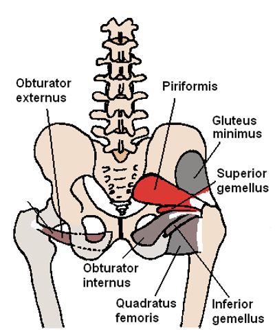
The piriformis is a muscle in the gluteal region of the lower limb. It is one of the six muscles in the lateral rotator group, BUT is one of the easiest to palpate.
Action: External rotator of the thigh
Attachments
Insertion Greater trochanter (leg bone)
Nickname
Pirif
Why is this important?
(1) Years of experience has taught us this usually a KEY muscle in solving back, hip and leg problems
(2) This is the closest hip muscle to the sciatic nerve. Please see the Tool box for therapy training.
(3) You will press THROUGH the Glute Max to work on the Piriformis

3. Gluteus medius
Action
The gluteus medius one of the three gluteal muscles, is a broad, thick, radiating muscle, situated on the outer surface of the pelvis
Actions (1)abduction of the hip;
(2) preventing adduction of the hip.
(3) Medial/internal rotation and flexion of the hip (anterior fibers).
(4) Extension and Lateral/external rotation of the hip (posterior fibers)
Attachments
Origin Gluteal surface of ilium, under gluteus maximus
Insertion Greater trochanter of the femur
Nickname
Glute med
Why is this important?
You'll usually discover this during a session as you feel the hip muscles You'll learn in the Tool box how to identify these "smaller" hip muscles.

4. Gluteus minimus
Beth ohara / CC BY-SA (http://creativecommons.org/licenses/by-sa/3.0/)
Action
The gluteus minimus, the smallest of the three gluteal muscles, is situated immediately beneath the gluteus medius
Actions: Works in concert with gluteus medius: 1) abduction of the hip, preventing adduction of the hip. 2) Medial rotation of thigh.
Attachments
Origin: From area in between the anterior gluteal line and inferior gluteal line of Gluteal surface ilium, under gluteus medius. (see photo)
Insertion: Greater trochanter of the femur
Nickname
Glute min
Why is this important?
If you learn to get good client feedback, you can note which muscles are key(s) during your therapy session. This will really help with follow up therapy and client enrollment.

5. Tensor Fasciae Latae
Beth ohara / CC BY-SA (http://creativecommons.org/licenses/by-sa/3.0/)
Action
The tensor fasciae latae is a muscle of the thigh. It is related with the gluteus maximus in function and structure and is continuous with the iliotibial tract, which attaches to the tibia. The muscle assists in keeping the balance of the pelvis while standing, walking, or running. I have heard it described as "Keeping the knee from buckling when the leg is extended.
Actions:
(1) Hip - flexion, medial rotation, abduction, (2) knee - lateral rotation, (3)Torso - stabilization
Attachments
Origin Anterior superior iliac spine
Insertion Iliotibial tract -The I.T. Band (via greater trochanter)
** You can see the TFL Starts at the curve in the pelvis, INTO the i t band, which continues down to the lateral knee**
Nickname
TFL
Why is this important?
This muscle is VERY important for athletes! One of the anatomy books says the TFL "helps keep the knee from buckling when the leg is extended." Our Tool Box will show you how to help loosen the TFL!
upper leg

6. Iliotibial band (I.T. Band)
Healthimage / CC BY-SA (https://creativecommons.org/licenses/by-sa/4.0)
Function
The iliotibial tract or iliotibial band is a longitudinal fibrous reinforcement of the fascia lata. The function/action of the ITB and its associated muscles is to extend, abduct, and laterally rotate the hip. In addition, the ITB contributes to lateral knee stabilization
Attachments
Origin: Anterolateral iliac tubercle portion of the external lip of the iliac crest
Insertion: Lateral condyle of the tibia
Nickname
i t band
Why is this important?
In the Tool Box, we will show you which muscles to work (and which angles to use!)

7. Biceps femoris (hamstring)
derivative work: r@ge (talk)Músculo_semimembranoso.png: *derivative work: r@geGluteus_maximus.png: Nikai / CC BY-SA (http://creativecommons.org/licenses/by-sa/3.0/)
Action
The biceps femoris is a muscle of the thigh located to the posterior, or back. As its name implies, it has two parts, one of which forms part of the hamstrings muscle group
Actions:
1) knee flexion, 2) internal and external rotation, and 3) hip extension
Attachments
Origin: a) long head: ischial tuberosity (lower pelvis) b) short head: proximal femur
Insertion: proximal fibula ** crosses two joints, the hip and the knee**
Nickname
biceps fem
Why is this important?
Tight hamstrings can affect the immediate area, plus the lower leg, PLUS back discomfort. We will explain more in the Tool Box page and Secret Sauce page.

8. Semimembranosis (hamstring)
BruceBlaus / CC BY-SA (https://creativecommons.org/licenses/by-sa/4.0)
Action
Attachments
Origin Ischial tuberosity (lower hip)
Insertion Medial condyle of tibia (lower leg)
Nickname
Semi memb
Why is this important?
Knowing the exact location of the three hamstring muscles will help you target your Neuromuscular Therapy AND help you have accurate notes - this will tell your client they are with the right therapist!

9. Semitendenosis (hamstring)
BruceBlaus / CC BY-SA (https://creativecommons.org/licenses/by-sa/4.0)
Action
Attachments
Origin Ischial tuberosity (lower hip)
Insertion Medial condyle of tibia (lower leg)
Nickname
semi tend
Why is this important?
Knowing the exact location of the three hamstring muscles will help you target your Neuromuscular Therapy AND help you have accurate notes - this will tell your client they are with the right therapist!
lower leg

10. Plantaris
Polygon data is generated by Database Center for Life Science (DBCLS) / CC BY-SA 2.1 JP (https://creativecommons.org/licenses/by-sa/2.1/jp/deed.en)
Action
The plantaris is one of the superficial muscles of the superficial posterior compartment of the leg (behind the knee). It is composed of a thin muscle belly and a long thin tendon. While not as thick as the achilles tendon, the plantaris tendon is the longest tendon in the human body
Actions: 1) Flexes foot and 2)Flexes knee
Attachments
Origin: Lateral supracondylar ridge of femur above lateral head of gastrocnemius (Distal lateral femur above knee)
Insertion: Tendo calcaneus - medial side, deep to gastrocnemius tendon (Inside of heel)
Nickname
plantaris
Why is this important?
Tightness of this muscle becomes noticeable with people who run FAST (track athletes, especially)
PS When working on a client who trains on the track, you will usually find the left plantaris to be tighter, because of the turns when running on a track.

11. Popliteus
Polygon data were generated by Database Center for Life Science (DBCLS)[2] / CC BY-SA 2.1 JP (https://creativecommons.org/licenses/by-sa/2.1/jp/deed.en)
Action
The popliteus muscle in the leg is used for (1) unlocking the knees when walking, by laterally rotating the femur on the tibia during the closed chain portion of the gait cycle. In open chain movements, the popliteus muscle(2) medially rotates the tibia on the femur. It is also used (3) when sitting down and standing up. (**note**) It is the only muscle in the posterior compartment of the lower leg that acts just on the knee and not on the ankle. The gastrocnemius muscle acts on both joints
Actions: 1) Medially rotates tibia on the femur if the femur is fixed (sitting down) or
2) Laterally rotates femur on the tibia if tibia is fixed (standing up),
3) Unlocks the knee to allow flexion (bending),
4) Helps to prevent the forward dislocation of the femur while crouching
Attachments
Origin: lateral femoral epicondyle (outside of leg just above the knee)
Insertion: posterior surface of tibia proximal to soleus line (inside front of lower leg)
Nickname
poplit
Why is this important?
You will want to palpate behind the knee to determine if this muscle is tight.
calves, primary

12. Gastrocnemus
Harrygouvas / Attribution
Action
The gastrocnemius muscle is a superficial two-headed muscle that is in the back part of the lower leg of humans. It runs from its two heads just above the knee to the heel, a three joint muscle.
Actions: (1) plantar flexes foot,
(2) flexes knee
Attachments
The tendons are in yellow!
The gastrocnemius muscle is named via Latin, from Greek γαστήρ "belly or stomach" and κνήμη "leg"; meaning "stomach of leg".
Origin: superior to articular surfaces of lateral condyle of femur and medial condyle of femur (both outside and inside of upper leg)
Insertion: tendo calcaneus (achilles tendon) into mid-posterior calcaneus (heel)
Nickname
Gastroc
Why is this important?
You'll want to palpate AND get client feedback to determine which lower leg muscle(s) are causing a concern.

13. Soleus
Polygon data is generated by Database Center for Life Science (DBCLS) / CC BY-SA 2.1 JP (https://creativecommons.org/licenses/by-sa/2.1/jp/deed.en)
Action
In humans and some other mammals, the soleus is a powerful muscle in the back part of the lower leg. It runs from just below the knee to the heel, and is involved in standing and walking.
Action: plantarflexion
NOTE: The Soleus does NOT flex the knee (but the Gastrocnemus and Plantaris do)
Attachments
Origin: fibula, medial border of tibia (both lower leg bones)
Insertion: tendo calcaneus (Achilles tendon)
Nickname
soleus
Why is this important?
You'll want to determine which lower leg muscle is causing a concern. Since the soleus is lower in the calf than the gasctroc, you should be able to feel if this is the "target" muscle for therapy.
Calves, stabilizers

14. Posterior Tibialis
Polygon data were generated by Database Center for Life Science (DBCLS)[2] / CC BY-SA 2.1 JP (https://creativecommons.org/licenses/by-sa/2.1/jp/deed.en)
Acton
The tibialis posterior is the most central of all the leg muscles, and is located in the deep posterior compartment of the leg
Actions:
(1) Inversion of the foot and
(2) plantar flexion of the foot at the ankle
Attachments
Origins: Tibia and fibula (The shin area)
Insertion: Navicular and medial cuneiform bone (Under foot)
Nickname
Post Tib
Why is this important?
When you work on the Posterior Tibialis, you'll probably also work on the Flexor hallucis longus muscle and the Flexor digitorum longus muscle which are in the same area, and also attach under the foot. These muscles are very important to athletic clients.

15. Peronius longus (and peronius brevis)
Henry Vandyke Carter / Public domain
Action
In human anatomy, the peroneus longus is a superficial muscle in the lateral (outside) compartment of the lower leg.
Actions: (1) plantarflexion, (2) eversion, (3) support arches
(Helps move and stabilize the foot)
Attachments
Origin: Proximal part of lateral surface of shaft of fibula (outside upper part of lower leg bone)
Insertion: First metatarsal, medial cuneiform (Attaches under the foot like a stirrup)
Nickname
Peron Long
Why is this important?
The peroneus longus and brevis are VERY important for people with ankle discomfort. The good news is they usually respond quickly to Neuromuscular Massage. I once worked on a Pole Vaulter during the U.S. Track trials and he was able to jump high enough to make the USA team.
bottom of foot

16. Plantar area
English wikipedia user Kosigrim / Public domain
Actions
There are 28 bones in the foot and about 33 muscles. This graphic only shows the percentage of areas where people feel pain.
Attachments
For our purposes, the important attachments are the tendon insertions of the muscles in the lower leg.
Nickname
Plantar
(If you find some tightness in a client's foot you can add the general location of tightness, which will help you focus on the correct part of the foot.)
Why is this important?
Our experience is that the large muscles are usually the culprit when solving tight muscle problems. If a client points to a certain part of the bottom foot, that will help you determine which muscle(s) to work on. We'll have more training in the Body Engineering page of "Secret Sauce".
achilles tendon

17. Achilles Tendon
Polygon data is generated by Database Center for Life Science (DBCLS) / CC BY-SA 2.1 JP (https://creativecommons.org/licenses/by-sa/2.1/jp/deed.en)
Function
Acting via the Achilles tendon, the gastrocnemius and soleus muscles cause plantar flexion of the foot at the ankle. This action brings the sole of the foot closer to the back of the leg. The gastrocnemius also flexes the leg at the knee.
Attachments
The Achilles is a tendon at the back of the lower leg, and is the thickest in the human body.[1][2][3][4][5][6][7] It serves to attach the plantaris, gastrocnemius (calf) and soleus muscles to the calcaneus (heel) bone. These muscles, acting via the tendon, cause plantar flexion of the foot at the ankle joint, and (except the soleus) flexion at the knee.
Nickname
Achilles
Why is this important?
Why? The Achilles does not "move" but it is acted upon by the lower leg muscles. If a client points to the Achilles tendon, that will indicate which muscle(s) you work on. While I rarely apply deep pressure on the Achilles tendon, clients usually enjoy some light cross friction in this area.
However, this can be a sensitive area, so you'll want to stay in communication.
Next - Muscle Box 4 - See link below
- Index
- About Us
- Testimonials
- How to start
- Business Box
- Muscle Box 1
- Muscle Box 2
- Muscle Box 3
- Muscle box 4
- Nerve Box
- Tool Box
- Reflexology
- Stretches
- Personal Development
- Finances
- Physical fitness
- Nutrition & Skin care
- Basic Home Therapy
- Advanced Subscriptions
- CEUs
- Live seminars
- Client Intake
- Body engineering
- Lessons we learned
- Business success
- Real Therapy videos