learn unique, effective techniques, and how to create too much business!
muscle box 2 - arms & hands
More muscles to learn!
You could search in Internet, read blogs, and watch lots of online videos, OR you can take a short cut and get some very good anatomy education RIGHT HERE!. We have done a LOT of leg work to put this valuable information in one place. PLUS, this information comes from many years of getting results for thousands of clients.
The "Muscle Box" - This is a list of muscles we learned to target as we perform therapy. If you learn this list, you'll have a good basic understanding of many of the tight muscles that can affect your clients. We have been getting performance feedback from clients since 1989 so we know this list is a vital key to your capability to help your clients.
You will see:
(a) The muscle name and picture,
(b) The action of the muscle
(c) The attachment points (We sometimes use generic descriptions.) and
(d) A "nickname" you can you use in your progress notes.
(e) Why is this important section. Information that can help you provide better results for your client.
Note: Even though this course is "basic anatomy", it is the building block of both your ability to perform our therapy style, AND your ability to communicate what you are doing.

John at Houston's Memorial Park
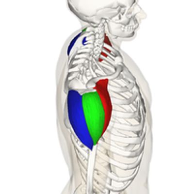
posterior arm

1. Rear Deltoid
Anatomography [CC BY-SA 2.1 jp (https://creativecommons.org/licenses/by-sa/2.1/jp/deed.en)]
Action
The deltoid muscle is the muscle forming the rounded contour of the human shoulder
Actions: shoulder abduction, flexion and extension
Attachments
Anterior border and upper surface of the lateral third of the clavicle , acromion and spine of scapula TO the upper arm (outside the collar bone, plus front of scapula to the upper arm)
Nickname
Delt (note: We know there are three Deltoids, but you'll know which Delt based on body position)
Why is this important?
Each area of concern has certain contributing tight muscles. As you learn our system and therapy protocols, you'll be better able to get results your clients can feel.

Action
Actions: a) Extend forearm, b)extend and adduct arm, c) extend shoulder
Attachments
Origins: a) front aspect of scapula (long head) and b) upper rear arm (lateral and medial head)
Insertion: the Olecranon process of Ulna (below elbow)
Nickname
triceps
Why is this important?
a) Muscle tightness concerns
b) Localized nerve impingement - SEE the Nerve Box page

3. Forearm extensors - Extensor digitorum
Mikael Häggström.When using this image in external works, it may be cited as:Häggström, Mikael (2014). "Medical gallery of Mikael Häggström 2014". WikiJournal of Medicine 1 (2). DOI:10.15347/wjm/2014.008. ISSN 2002-4436. Public Domain.orBy Mikael Häggström, used with permission. [Public domain]
Action
The extensor digitorum muscle is a muscle of the posterior forearm. It extends the medial four digits of the hand. It is innervated by the posterior interosseous nerve, which is a branch of the radial nerve
Actions: Extend hand, wrist, fingers
Attachments
Origin: Lateral epicondyle (elbow)
Insertion: 2nd, 3rd, 4th & 5th fingers
Nickname
Ext digit
Why is this important?
We have found this to be very important for BOTH tennis elbow tightness and carpal area tightness. We will teach you several therapy techniques in the Tool Box.

4. Extensor carpi ulnaris
By Mikael Häggström, used with permission. - File:Gray418.png
Action
The extensor carpi ulnaris is a skeletal muscle located on the ulnar side of the forearm. It acts to extend and adduct at the carpus/wrist.
Action: Extend and adduct the wrist.
Attachments
Origin: Lateral epicondyle of the Humerus & Olecranon of Ulnar (elbow)
Insertion: 5th metacarpal (hand)
Nickname
Forearm (simple, but you'll know which muscle(s) by the feel.
Why is this important?
This is one of SEVERAL important muscles in the forearm (note the drawing). We don't feel you need to "memorize" each name, but it is important to learn to palpate and work on whichever extensor muscle you find to be tight.

5. Supinator
By Henry Vandyke Carter - Henry Gray (1918) ) Gray's Anatomy, Plate 420, Public Domain, https://commons.wikimedia.org/w/index.php?curid=552307
Action
The supinator is a broad muscle in the posterior compartment of the forearm, curved around the upper third of the radius. Its function is to supinate (rotate the palm upward) the forearm
Action: Rotation of the forearm and hand so that the palm faces forward or upward
Attachments
Origin: Lateral epicondyle of humerus & supinator crest of ulna (elbow + lower arm bone)
Insertion: Lateral Radius (outside of other lower arm bone)
Nickname
Supinator
Why is this important?
This is a good example of the importance of a thorough new client interview. We used to call this the "ping pong" muscle because of it's function in that sport. If you learn what people do, or what type of injury they have had, you will learn "patterns of tightness" that will guide you where to work. We will teach you more in the Tool Box and in the Secret Sauce pages.

Action
It is partly blended in with the triceps, which it (1) assists in extension of the forearm. It also (2) stabilizes the elbow during pronation and supination and (3) pulls slack out of the elbow joint capsule during extension to prevent impingement. ALSO IMPORTANT with Extensor Carpi (see above)
Attachments
Origin: lateral epicondyle of the humerus (elbow)
Insertions: Lateral radius AND posterior Ulna (both bones of the lower arm)
Nickname
Anconeus
Why is this important?
This is one of many examples of the importance of "smaller" muscles. They often try to help the larger muscle in an area, and then get overworked or tight. We will show you how to identify this muscle through the client interview process or by palpation.
anterior arm (and chest)

7. Pectoralis Major
CC BY-SA 3.0, https://commons.wikimedia.org/w/index.php?curid=239265
Action
The pectoralis major is a thick, fan-shaped muscle, situated at the chest of the human body. It makes up the bulk of the chest muscles and lies under the breast.
Actions: 1) Clavicular head: flex the humerus
2) Sternocostal head: horizontal and vertical adduction, extension, and internal rotation of the humerus
Attachments
Origins: a) anterior clavicle b) anterior sternum, SIX costal cartilidges, c) the external oblique
Insertion: humerus
Nickname
Pect
Why us this important?
a) This is a good example of finding tightness in one area and discomfort in a different area
b) We have found several ways to release this area. Please look in the Tool Box page and the Secret Sauce page.

8. Anterior Deltoid
The part in red: Anatomography / CC BY-SA 2.1 JP (https://creativecommons.org/licenses/by-sa/2.1/jp/deed.en)
Action
Attachments
Nickname
Delt (You will know which Delt you are working on by seeing the client's chart.)
Why is this important?
This is one of several muscles that are difficult to reach based on standard massage positioning. We will teach you effective ways to reach these types of muscles.

9. Biceps
Anatomography / CC BY-SA 2.1 JP (https://creativecommons.org/licenses/by-sa/2.1/jp/deed.en)
Action
The biceps is a large muscle that lies on the front of the upper arm between the shoulder and the elbow. Both heads of the muscle arise on the scapula and join to form a single muscle belly which is attached to the upper forearm
Actions:
2) flexes and abducts shoulder
3) supinates radioulnar joint in the forearm
Attachments
Origins: Short head: coracoid process ("hooked" edge of shoulder blade) of the scapula.
Long head: supraglenoid tubercle (part of shoulder blade where the arm connects)
Insertions: Radial tuberosity and bicipital aponeurosis into deep fascia on medial part of forearm
Nickname
Biceps
Why is this important?
The biceps can be VERY achy (for the client) to work on. In the Tool Box we will teach you several effective therapy techniques that stay within the client's comfort zone.
other muscles in the upper arm

10. Coracobrachialis
Anatomography / CC BY-SA 2.1 JP (https://creativecommons.org/licenses/by-sa/2.1/jp/deed.en)
Action
Attachments
Origin Coracoid process of scapula
Insertion Anteromedial surface of humerus distal to crest of lesser tubercle (Distal aspect of humerus, but above the elbow)
Nickname
coracobrachialis
Why is this important?
You should be able to palpate this muscle as you work on the upper arm, especially if the biceps is NOT the culprit muscle.

11. Brachialis
By Anatomography - , CC BY-SA 2.1 jp, https://commons.wikimedia.org/w/index.php?curid=27462870
Action
The brachialis is a muscle in the upper arm that flexes the elbow joint. It lies deeper than the biceps brachii. The brachialis is the prime mover of elbow flexion (see below).
Action: flexion at elbow joint
Attachments
Origin anterior surface of the humerus, particularly the distal half of this bone (mid part of upper arm)
Insertion proximal aspect of the ulna (inside bone of lower arm)
Nickname
brachialis
Why is this important?
While the biceps brachii appears as a large anterior bulge on the arm and commands considerable interest among body builders, the brachialis underlying it actually generates about 50% more power and is thus the prime mover of elbow flexion
Forearm muscles

12. Forearm Brachioradialis
Henry Vandyke Carter / Public domain
Action(s)
The brachioradialis is a muscle of the forearm that flexes the forearm at the elbow. It is also capable of both pronation (rotate palm downward) and supination (rotate palm upward), depending on the position of the forearm.
Actions (1) Flexion of elbow, (2) supination and(3) pronation of the radioulnar joint to 90°
Attachments
Origin Lateral supracondylar ridge of the humerus (Above elbow)
Insertion Distal radius (radial styloid process) (Close to wrist)
Nickname(s)
brachio (You'll know the full name based on which body part you are working on -the forearm)
Why is this important?
Client feedback has taught us that this is a KEY muscle in helping with tennis/golf elbow AND carpal tunnel tightness. We will teach more in the Tool Box page and the Secret Sauce page.

13. Several forearm flexors, i.e. Flexor carpi radialis
Henry Vandyke Carter / Public domain
Action
The flexor carpi radialis is a muscle of the human forearm that acts to flex and abduct the hand. The Latin carpus means wrist; hence flexor carpi is a flexor of the wrist.
Action: Flexion of the wrist.
Attachments
Origin Medial epicondyle of humerus (elbow)
Insertion Bases of second and third metacarpals (hand)
Nickname
flexors
Why is this important?
There are a number of flexors (and extensors on the front of the forearm). As you work on clients, you might find one or more of these "smaller" muscles to be tight. We will share more information in the Tool Box section.
"other" arm muscles

14. Other arm muscles
Henry Vandyke Carter / Public domain
Action
Flexion of the wrist and/or fingers
Attachments
Origins: Usually the humerus
Insertions: The radius (wrist) or fingers
Nickname
"medial" or "lateral" flexor (This will depend on what you find to be tight during therapy.)
Nickname
medial (or lateral) flexor
Why is this important?
This is really a way for you to notate the location of the tight "small" muscles you find during therapy. I had one client who does needle point stitching, so we identified just one of the small muscles during her session. This will help you keep track of what you find. Your clients will LIKE the fact that you kept notes!

15. Thumb adductor - Adductor pollicis
KenhubTemplate:Author of Illustration : Yousun Koh / CC BY (https://creativecommons.org/licenses/by/3.0)
Action
The adductor pollicis muscle is a muscle in the hand that functions to adduct the thumb. It has two heads: transverse and oblique. It is a fleshy, flat, triangular, and fan-shaped muscle deep in the hand at the center of the palm.
Action: Adduct (move inward) the thumb
Attachments
Origins: 2nd and 3rd metacarpals (hand)
Insertion: Medial aspect of thumb
Nickname
Thumb (this is just the location of the therapy pressure). We will teach you at least two ways to work on this muscle/area.
Why is this important?
Most of the "major player" hand muscles are not in the hand; rather the forearm. (So we rarely do deep pressure in the hand itself.)The adductor polllicis is an exception, and we will show you two ways you can work on it.
hands

16. Hands
KenhubTemplate:Author of Illustration : Yousun Koh / CC BY (https://creativecommons.org/licenses/by/3.0)
Actions
Flexion and abduction of the fingers, and adduction of the thumb
Attachments
Various carpels and metacarpals in the hand
Nickname
Your note will vary according to what you (and the client) find to be tight: (i.e. lateral, medial, small finger, thumb)
Why is this important?
One of my jokes is: "Size matters." The smaller muscles in the body are usually not the major contributor to the client's concern. In the hand, the thumb adductor is an exception. However, sometimes the these smaller muscles can be important. Be sure you note your finding.
Also it might feel very good to your client if you kneed or massage their hands.
Next - Muscle Box 3 - See link below
- Index
- About Us
- Testimonials
- How to start
- Business Box
- Muscle Box 1
- Muscle Box 2
- Muscle Box 3
- Muscle box 4
- Nerve Box
- Tool Box
- Reflexology
- Stretches
- Personal Development
- Finances
- Physical fitness
- Nutrition & Skin care
- Basic Home Therapy
- Advanced Subscriptions
- CEUs
- Live seminars
- Client Intake
- Body engineering
- Lessons we learned
- Business success
- Real Therapy videos